
3D Bioprinted Organoids for Drug Screening are changing the way researchers study human health and disease. Traditional laboratory models often fail to capture the complexity of human tissues. As a result, researchers are increasingly turning to advanced 3D models that better mimic real biological environments.
In pregnancy research, this technology offers exciting opportunities. Scientists can now create miniature tissue models that closely resemble the placenta and other organs involved in maternal and fetal health. These models help researchers understand disease mechanisms, identify biomarkers, and evaluate potential therapies before they reach clinical trials.
Recent advances in bioprinting and organoid technology are improving our ability to study pregnancy complications, including preeclampsia, while reducing dependence on animal models.
What Are 3D Bioprinted Organoids?
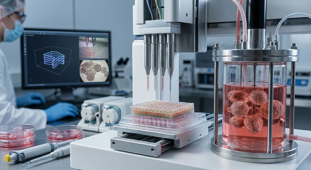
Organoids are miniature three-dimensional structures grown from cells that replicate key features of real organs. When combined with bioprinting technology, researchers can precisely arrange cells and biomaterials to create highly realistic tissue models.
Unlike traditional cell cultures, organoids provide the following:
- More realistic cell interactions
- Better tissue organization
- Improved biological function
- Enhanced disease modeling capabilities
These advantages make organoids valuable tools for biomedical research and drug development.
Understanding Bioprinting Technology
Bioprinting uses specialized printers to deposit living cells and biomaterials layer by layer.
Researchers can control:
- Cell placement
- Tissue architecture
- Structural support materials
- Microenvironment conditions
This precision allows scientists to recreate complex biological systems that closely resemble human tissues.
The technology has become increasingly important in reproductive and placental research.
Why Traditional Drug Screening Models Have Limitations
Drug development often relies on animal models and two-dimensional cell cultures.
While useful, these approaches have several limitations:
- They may not accurately reflect human biology.
- Cell behavior differs in flat cultures.
- Animal responses may not match human responses.
- Important tissue interactions can be missed.
These limitations contribute to high failure rates during clinical development.
3D bioprinted organoids help bridge this gap by providing more physiologically relevant models.
Placental Organoids and Pregnancy Research
The placenta plays a critical role during pregnancy. It regulates nutrient exchange, oxygen delivery, hormone production, and immune interactions between mother and baby.
Because access to living placental tissue during pregnancy is limited, researchers have sought alternative models.
Placental organoids provide a powerful solution.
Recent studies have demonstrated how organoids can mimic important aspects of placental development and function. These models enable researchers to investigate normal pregnancy processes as well as disease mechanisms.
Studying Early Placental Development
Research titled “Matrix Directs Trophoblast Differentiation in a Bioprinted Organoid Model of Early Placental Development” highlighted how the surrounding cellular environment influences placental cell behavior.
The study demonstrated that extracellular matrix components play a major role in trophoblast differentiation.
Trophoblast cells are essential for:
- Placental formation
- Maternal blood vessel remodeling
- Healthy fetal growth
Understanding these processes may help researchers identify new therapeutic targets for pregnancy complications.
Mini-Placentas and Disease Modeling
Recent advances have also led to the development of 3D printed mini-placentas.
These miniature tissue models provide researchers with an innovative platform for studying:
- Preeclampsia
- Placental insufficiency
- Fetal growth restriction
- Maternal vascular disorders
Mini-placentas allow scientists to observe biological processes that would otherwise be difficult to study in real-time.
They also create new opportunities for personalized medicine approaches.
Biomarker Discovery Using Placental Organoids
Biomarker discovery remains a major focus in pregnancy research.
Researchers use organoids to identify biological signals associated with disease progression.
Potential biomarkers may help:
- Predict pregnancy complications
- Improve early diagnosis
- Monitor treatment response
- Support risk stratification
Placental organoids offer a controlled environment where researchers can evaluate these biomarkers with greater accuracy.
As a result, they are becoming valuable tools for translational research.
Improving Drug Screening and Development
One of the most promising applications of organoid technology is drug screening.
Researchers can test candidate therapies directly on human-like tissues before clinical studies begin.
Benefits include:
- Faster identification of effective therapies
- Improved safety assessment
- Reduced development costs
- Better prediction of clinical outcomes
This approach may accelerate the development of treatments for conditions that currently have limited therapeutic options.
Applications in Preeclampsia Research
Preeclampsia remains one of the leading causes of maternal and fetal complications worldwide.
Researchers are increasingly using placental organoids to investigate:
- Angiogenic imbalance
- Oxidative stress
- Inflammatory signaling
- Trophoblast dysfunction
- Vascular abnormalities
These studies help reveal the biological pathways involved in disease progression.
They also provide platforms for testing novel therapies in a controlled environment.
Beyond the Placenta: Cardiovascular Health Models
The impact of pregnancy complications often extends beyond delivery.
Research using 3D in vitro cardiovascular models has revealed important insights into postpartum health following hypertensive disorders of pregnancy.
These models allow researchers to study:
- Long-term cardiovascular changes
- Cardiac remodeling
- Vascular dysfunction
- Disease-specific molecular signatures
Such findings may help explain why women with a history of preeclampsia face higher cardiovascular risks later in life.
3D Cardiac Spheroids and Preeclampsia
Researchers have also developed 3D cardiac spheroid models to investigate the cardiovascular consequences of preeclampsia.
These models mimic heart tissue behavior and provide a platform for studying:
- Cardiac stress responses
- Cellular remodeling
- Molecular signaling pathways
- Potential therapeutic interventions
Combining placental and cardiac models offers a more comprehensive understanding of maternal health.
Challenges and Future Opportunities
Despite significant progress, several challenges remain.
Researchers continue working to improve:
- Model complexity
- Long-term tissue stability
- Standardization
- Clinical translation
Future developments may integrate:
- Artificial intelligence
- Advanced imaging technologies
- Personalized patient-derived tissues
- Multi-organ systems
These innovations will further enhance the accuracy and usefulness of organoid-based research.
Conclusion
3D Bioprinted Organoids for Drug Screening are transforming modern biomedical research. By creating realistic models of placental and cardiovascular tissues, researchers can better understand disease mechanisms, identify biomarkers, and evaluate new therapies.
Studies involving bioprinted placental organoids, mini-placentas, and cardiac spheroids demonstrate the growing potential of this technology. As these models continue to evolve, they may accelerate drug development, improve pregnancy outcomes, and advance personalized medicine for women’s health.